
Digital Dental X-Ray & 3D Imaging
Ultra-low radiation digital sensors and high-definition panoramic imaging for crystal-clear diagnoses and painless treatment planning.
What is a Digital Dental X-Ray?
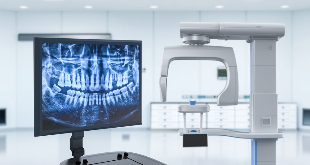
A digital dental X-ray uses high-sensitivity electronic sensors instead of conventional photographic film to capture high-definition views of your teeth, roots, and jawbone. The images appear instantly on our clinic monitors with zero chemical processing time.
Seeing Beyond the Surface
Over 50% of dental pathology — such as root canal infections, bone resorption, impacted wisdom teeth, and interproximal cavities — is hidden below the gumline and invisible to the naked eye. Digital X-rays allow us to spot issues before they cause pain.
When Do You Need a Dental X-Ray?
Digital imaging is an essential diagnostic tool for various clinical needs, including:
Routine Annual Screenings
Monitor bone levels and spot hidden interdental decay early.
Sudden Toothache or Trauma
Locate root fractures, abscesses, or deep nerve inflammation.
Implants, Ortho & Root Canals
Precise measurement of canal depth, bone thickness, and tooth angles.
What Can Digital X-Rays Detect?
Hidden Interdental Cavities
Decay forming between tight teeth that is completely missed on visual exams.
Root Tip Abscess & Infection
Nerve necrosis and periapical lesions requiring immediate root canal care.
Jawbone Resorption
Evaluating bone density lost from advanced periodontal (gum) disease.
Impacted Wisdom Teeth
Mapping tooth roots relative to nerves before surgical extraction.
Recurrent Decay Under Crowns
Identifying leakage or decay beneath existing fillings and bridges.
Implant Site Planning
Precise height and width mapping for permanent dental implant placement.
Types of Digital X-Rays
Periapical (IOPA) X-Ray
Captures the entire tooth from crown to root tip and surrounding bone. Crucial for root canals and deep decay.
Bitewing X-Ray
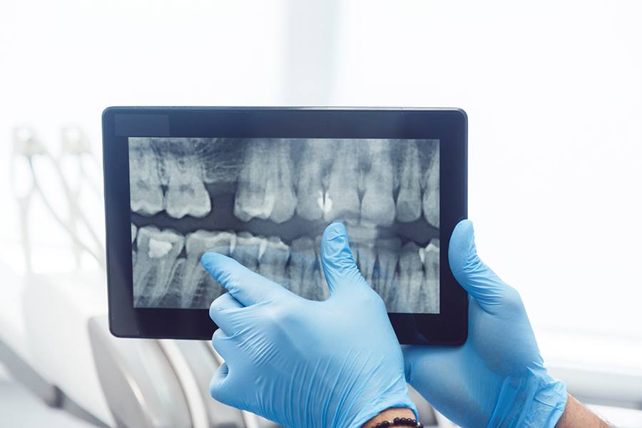
Shows upper and lower molars in one view to detect cavities forming between tight teeth and check bone height.
Panoramic OPG X-Ray
A broad 2D scan capturing your full upper and lower jaw, sinuses, and TMJ joints in a single rotation.
Why Digital X-Rays Are Safe & Superior
Ultra-Low Radiation
Digital sensors require up to 90% less radiation than traditional film, making it as safe as a short outdoor stroll.
Instant Display
No chemical developing wait. High-resolution images appear on our chairside monitors within 3 seconds.
HD Contrast Enhancement
Doctors can zoom in, adjust exposure, and colorize pixels to detect micro-cracks before they expand.
Full Lead Apron Shielding
We provide protective lead aprons with thyroid collars for every single patient as standard safety protocol.
Our Step-by-Step Imaging Process
Fast, completely painless, and finished in under 2 minutes:
Clinical Screening
Dentist evaluates which specific views are needed.
Protective Shielding
Lead apron and thyroid collar placed for safety.
Sensor Placement
Soft, comfortable digital sensor placed next to tooth.
Instant Capture
Image takes 0.2 seconds and appears on monitor.
Chairside Review
Doctor explains the findings directly on the screen.
Tailored Treatment
Clear, transparent diagnosis and treatment plan.
Digital Imaging Charges
100% Transparent & Fair Pricing
Intraoral Digital X-Ray (IOPA)
Periapical single tooth high-resolution scan
Panoramic X-Ray (OPG)
Full-mouth panoramic scan for ortho & surgery
* Note: The exact view required will be determined by your dental specialist during examination to ensure the lowest exposure and maximum clinical accuracy.
Book Digital DiagnosticDiagnostic Imaging Gallery
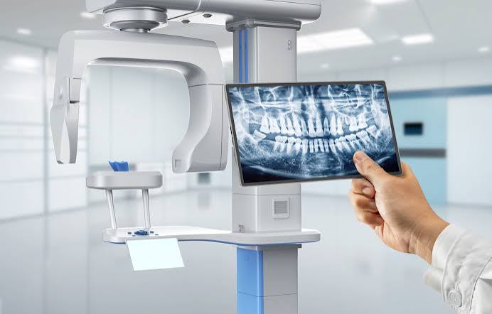
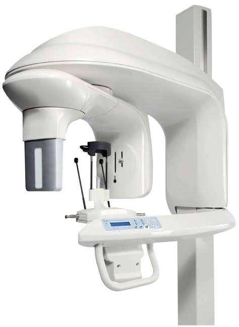
Explore the modern equipment and chairside monitors we use to ensure your treatments are accurate and painless.
Fast & Painless Diagnostics
“I had an agonizing toothache with no visible cavity on the outside. The doctor took a digital X-ray in under 10 seconds and showed me the deep root infection on the screen. The speed and clarity allowed us to start treatment immediately with zero delay.”
Frequently Asked Questions
Related Dental Services


Accurate Diagnosis Leads to Lasting Oral Health
Experiencing tooth pain or need a comprehensive dental check-up? Book your digital diagnostic evaluation today.